Ultrasound scan
Peer reviewed by Dr Hayley Willacy, FRCGP Last updated by Dr Toni Hazell, FRCGPLast updated 19 Sept 2023
Meets Patient’s editorial guidelines
- DownloadDownload
- Share
- Language
- Discussion
- Audio Version
- Add to preferred sources on Google
An ultrasound scan is a painless test that uses sound waves to create images of organs and structures inside your body. It is a very commonly used test. As it uses sound waves it is thought to be very safe.
Doppler and duplex scans are used to visualise blood or fluids flowing through the body.
Note: the information below is a general guide only. The arrangements, and the way tests are performed, may vary between different hospitals. Always follow the instructions given by your doctor or local hospital.
At a glance
An ultrasound scan uses high-frequency sound waves to create images of inside your body.
It is a safe and painless test that does not use radiation.
Ultrasound scans can help monitor unborn babies, detect organ problems, or examine muscles and joints.
Specific types of ultrasound include Doppler for blood flow and duplex for combined imaging.
You might need to prepare for some scans, for example by drinking fluid to fill your bladder.
After the scan, a report is sent to the doctor who requested it, who will discuss the results with you.
Sign up for our free 8-week Healthy Pregnancy course!
Each week we’ll share useful information and essential tips on topics such as nutrition, exercise, mental health, symptoms to look out for, and preparing for childbirth, to help you navigate your pregnancy journey whatever stage you are at.
By subscribing you accept our Privacy Policy. You can unsubscribe at any time. We never sell your data.
What is an ultrasound scan?
An ultrasound scan is a safe and painless test that creates images of organs, glands, abnormal lumps and other structures like muscles, tendons and joints, as well as being used to monitor the growth and development of the fetus in pregnancy.
What is ultrasound?
Ultrasound is a high-frequency sound that you cannot hear but it can be emitted and detected by special machines.
What is an ultrasound used for?
It is used in many situations. The way the ultrasound bounces back from different tissues can help to determine the size, shape and consistency of organs, structures and abnormalities. So it can:
Help to monitor the growth of an unborn child and check for abnormalities. An ultrasound scan is routine for pregnant women.
Detect abnormalities of heart structures such as the heart valves. This type of ultrasound scan is called echocardiography. See the separate leaflet called Echocardiogram for more details.
Help to diagnose problems of internal organs such as the:
Liver.
Gallbladder.
Pancreas.
Thyroid gland.
Lymph nodes.
Ovaries.
Testes.
Kidneys.
Bladder.
Appendix.
For example, it can help to determine if an abnormal lump in one of these organs is a solid tumour or a fluid-filled cyst. Ultrasound also helps look for stones in the gallbladder or kidney.
Help determine the nature of breast lumps. Ultrasound is one of the tests used to establish if a lump is non-cancerous (benign) or breast cancer.
Help diagnose problems with muscles, tendons and joints. For example, ultrasound scans are used to help diagnose:
Detect abnormal widening of blood vessels (aneurysms).
Guide internal biopsies. A biopsy is a procedure in which a sample of tissue is taken. Some biopsies are taken using a thin needle, and the needle is guided to the right place with an ultrasound scan. For example, if you have a lump in your breast, you may have a sample of the lump taken away. The sample is then examined under the microscope to see if your lump is cancerous or not.
Check if something that has been placed into your body is still there. For example, if you use a contraceptive coil (intrauterine device) and the threads are not visible, a scan will tell you if the coil is still there or has fallen out.
Some specialist ultrasound techniques
In some situations, a clearer picture can be obtained from a probe that is within the body. So a small probe, still attached by a wire to the ultrasound machine, can be:
Swallowed into the gullet (oesophagus). This may be used to obtain clearer images of the internal organs, particularly the stomach, upper gut and pancreas.
Placed in the vagina or rectum to obtain clearer images of inner organs, such as the womb (uterus), ovaries or prostate gland.
Used to help guide a surgeon during an operation, in order to look deeper into structures.
Ultrasound may also be used for treating certain conditions, particularly those of muscles, tendons and joints. The scan may be used to guide an injection which can help to treat the problem.
Doing the injection with the help of an ultrasound scan makes sure it reaches exactly the right place. For example, ultrasound-guided injections are a common way to treat shoulder problems such as a frozen shoulder.
The above are not exhaustive lists, and ultrasound scanning has other uses.
Preparing for an ultrasound scan
Usually there is no special preparation needed. Continue to take your usual medication. You should eat and drink normally before and after the test unless otherwise instructed. For example:
If certain parts of the tummy (abdomen) are being examined, you may be asked to eat a low-fibre diet for a day or so before the test (to minimise 'gas' in your gut).
You may be asked not to eat for several hours before a scan of the abdomen.
Occasionally for some scans, you may be given an enema to clear the bowel.
To scan the bladder or pelvis, you may be asked to drink some fluid before the test so that you have a full bladder. This is particularly likely if you are having a scan in pregnancy, or a scan of your ovaries or womb (uterus).
If your renal tract (kidneys and bladder) are being scanned, you may be asked to attend with a full bladder, have the first scan, empty your bladder, and then be scanned again. This is to check if your bladder is fully emptying when you go to the toilet.
You will be told what you need to do before any particular scan.
How does ultrasound work?
Ultrasound travels freely through fluid and soft tissues. However, ultrasound bounces back (is reflected back) as echoes when it hits a more solid (dense) surface. For example, the ultrasound will travel freely though blood in a heart chamber. But, when it hits a solid valve, a lot of the ultrasound echoes back.
Another example is that when ultrasound travels though bile in a gallbladder it will echo back strongly if it hits a solid gallstone - as in the ultrasound image below. The arrow points to a gallstone in the gallbladder.
Gallstone ultrasound image
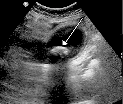
© By James Heilman (Own work), via Wikimedia Commons
So, as ultrasound 'hits' different structures of different density in the body, it sends back echoes of varying strength.
What does an ultrasound scan involve?
You lie on a couch and an operator places a probe on your skin over the part of your body to be examined. The probe is a bit like a very thick blunt pen. Lubricating jelly is put on your skin so that the probe makes good contact with your body. The image below shows an ultrasound scan of the neck.
Neck ultrasound scan
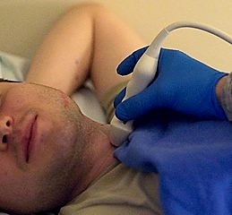
© By Senior Airman David C Danford, released [Public domain], via Wikimedia commons
The probe is connected by a wire to the ultrasound machine, which is linked to a monitor. Pulses of ultrasound are sent from the probe through the skin into your body. The ultrasound waves then bounce back as echoes from the various structures in the body.
The echoes are detected by the probe and are sent down the wire to the ultrasound machine.
Ultrasound scanning machine
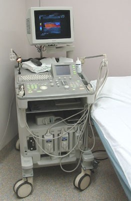
© By Daniel W Rickey, via Wikimedia Commons
They are displayed as a picture on the monitor. The picture is constantly updated so the scan can show movement as well as structure. For example, the valves of a heart opening and closing during a scan of the heart.
The operator moves the probe around over the surface of the skin to obtain views from different angles.
The scan is painless and takes about 15-45 minutes, depending on which parts of the body are being examined. A record of the results of the test can be made as still pictures or as a video recording, and a report summarising the finding will also be written.
What is a Doppler ultrasound scan?
A Doppler ultrasound records sound waves reflecting off moving objects, such as blood cells, to measure their speed and other aspects of how they flow through the body.
How does Doppler ultrasound work?
If the structure is moving then the echo comes back at a slightly different frequency (called the Doppler effect). This difference in frequency can be used to measure the speed of movement.
Blood moving in an artery or vein causes small echoes and these are used to measure the speed of movement of the blood cells. The sound waves may be amplified though speakers.
This allows the practitioner to listen to the flow of blood cells to determine whether or not there is normal flow. For example, listening to the flow of blood through the heart of a baby during a routine antenatal check-up.
The sound waves may also be converted to colour pictures on a screen so that flow can be seen through the arteries or veins (colour Doppler) - as below.
Colour Doppler ultrasonography
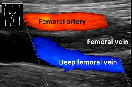
© By Mikael Häggström [CC0] (Own work), via Wikimedia Commons
They may also be plotted on a graph showing changes in speed and direction (velocity).
What is Doppler ultrasound used for?
To listen to the heartbeat of an unborn baby (fetus) during pregnancy.
To examine blood flow in arteries or veins in your arms or legs to see if you might have:
Injury to your veins or arteries following trauma.
What does a Doppler ultrasound involve?
During pregnancy, the Doppler ultrasound is very similar to an ultrasound scan. A probe covered with gel is put on your skin over the pregnant womb (uterus). This is connected to a speaker. You and the practitioner are able to listen to the flow of blood through the baby's heart.
During a Doppler ultrasound of the arms and legs, blood pressure cuffs are placed along the thigh, calf, or ankle, or to different points along the arm. A paste is applied to the skin over the arteries being examined. Images are created as the probe is moved over each area.
What is duplex ultrasound?
Duplex ultrasound is a special technique which combines traditional ultrasound with Doppler ultrasound. Images of the solid object being examined - for example, the artery and the blood flowing through it - are displayed on a screen or monitor.
The object is usually grey and the blood flow is usually in colour (colour Doppler).
What is duplex ultrasound used for?
Duplex ultrasound is most commonly used to evaluate the blood flow in various arteries and veins in the body. The scan can help diagnose the following conditions:
Widening of the main artery in the tummy (abdominal aortic aneurysm). Ultrasound scans are used in the national screening programmes across the UK for abdominal aortic aneurysm.
Blockage to an artery (an arterial occlusion).
Blood clot.
Blockage to the arteries in the neck (carotid occlusive disease).
Renal duplex examines the kidneys and their blood vessels.
Venous insufficiency (a condition where veins have a problem sending blood back to the heart).
The images below are produced from a Doppler scan of the kidney.
Kidney ultrasound scan
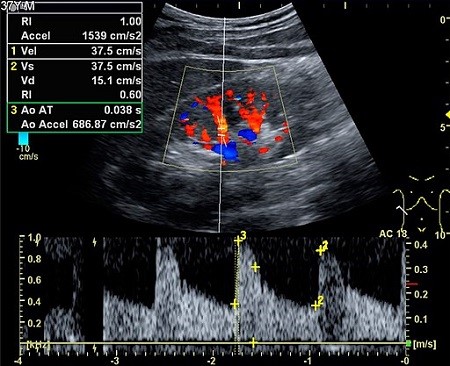
© By Kristoffer Lindskov Hansen, Michael Bachmann Nielsen and Caroline Ewertsen [CC BY 4.0 (https://creativecommons.org/licenses/by/4.0)], via Wikimedia Commons
What does a duplex ultrasound involve?
This test is very similar to an ultrasound scan. A probe covered with gel is placed over the area to be examined. Images of the solid organ and the blood flowing through it are then seen on a monitor.
Are there any side-effects?
These scans are painless and safe. Unlike X-rays and other imaging tests, they do not use radiation. They have not been found to cause any problems or complications.
What happens after an ultrasound scan?
The person operating the scanning machine will have put some jelly on to your skin at the start of the procedure. Once the scan is completed, this jelly will be wiped off and you can get dressed and leave.
The scan operator sometimes talks to you during the scan, so you may immediately have an idea of what was found. A report also needs to be written and this is sent to the person who ordered your scan - your GP or specialist team.
They will be able to discuss your result when you see them next. The results are normally available within a couple of weeks. It is important that you get the result from the team who requested your scan - so if the scan was requested by a hospital consultant, you should contact their secretary for the result, not your GP.
Patient picks for Imaging

Tests and investigations
CT colonography
CT stands for computed tomography. CT colonography uses a CT scanner to produce detailed pictures of the colon and rectum. This test can be used instead of a colonoscopy to help detect cancers and other bowel conditions. Note: the information below is a general guide only. The arrangements, and the way tests are performed, may vary between different hospitals. Always follow the instructions given by your doctor or local hospital.
by Dr Rachel Hudson, MRCGP
Tests and investigations
MRI scan
An MRI scan is a safe and painless test that can provide detailed pictures of organs and other structures inside your body. Note: the information below is a general guide only. The arrangements (and the way tests are performed) may vary between different hospitals. Always follow the instructions given by your doctor or local hospital. These are usually included with your appointment letter.
by Dr Toni Hazell, FRCGP
Frequently asked questions
Will an ultrasound scan be painful?
No, ultrasound scans are generally painless. You lie on a couch, and a probe with lubricating jelly is placed on your skin over the area to be examined. The process itself is not painful, and there are no known problems or complications associated with it.
How long does an ultrasound scan typically take?
The duration of an ultrasound scan can vary. It usually takes between 15 to 45 minutes, depending on which specific parts of the body are being examined.
What is the lubricating jelly used for during an ultrasound?
Lubricating jelly is applied to your skin so that the ultrasound probe can make good, continuous contact with your body. This allows the ultrasound waves to travel effectively through your skin and into your body, and for the echoes to be detected clearly.
Why would an internal ultrasound probe be used instead of an external one?
In some specific situations, an internal probe, placed within the body (like being swallowed or inserted into the vagina or rectum), can provide clearer images, especially for organs like the stomach, upper gut, pancreas, womb, ovaries, or prostate gland. This method allows for a more direct view of these inner structures.
Can an ultrasound scan be used to help treat certain conditions?
Yes, ultrasound can be used for treating certain conditions, particularly those affecting muscles, tendons, and joints. The scan may be used to guide an injection precisely to the problem area, ensuring it reaches the exact right place for effective treatment, such as for shoulder problems like a frozen shoulder.
How soon will I get the results of my ultrasound scan?
A report summarizing the findings of your scan will be written and sent to the person who requested it, such as your GP or specialist team. The results are normally available within a couple of weeks. It's important to get the results from the team who originally requested your scan.
Further reading and references
- Dietrich CF, Mathis G, Cui XW, et al; Ultrasound of the pleurae and lungs. Ultrasound Med Biol. 2015 Feb;41(2):351-65. doi: 10.1016/j.ultrasmedbio.2014.10.002.
- Guidelines for Professional Ultrasound Practice; Society and College of Radiographers and British Medical Ultrasound Society (SCoR/BMAS), 2019
- Ulrich CC, Dewald O; Pregnancy Ultrasound Evaluation.
About the authorView full bio

Dr Toni Hazell, FRCGP
MBBS, BSc, FRCGP, DFSRH, Dip GU med, DRCOG, DCH (London, UK, 2000)
Dr. Toni Hazell qualified from St. Mary’s Hospital Medical School and did her VTS at Northwick Park Hospital.
About the reviewerView full bio

Dr Hayley Willacy, FRCGP
General Practitioner, Medical Author
MBChB (1992), DRCOG, DFFP, MRCOG (Part 1) MRCGP (2007), DFSRH (2013), MSc - medical education (2020)
Dr Hayley Willacy was an NHS GP working in northwest England, who retired from clinical practice in 2022 after 30 years.
Article history
Article also available in English, German, Spanish, French, Italian, Portuguese, Hindi, Hebrew, Arabic, and Swedish.
Next review due: 17 Sept 2028
19 Sept 2023 | Latest version
Join the conversation
Share your thoughts or experiences with others in our patient communities.

Ask, share, connect.
Browse discussions, ask questions, and share experiences across hundreds of health topics.

Feeling unwell?
Assess your symptoms online for free
Sign up to the Patient newsletter
Your weekly dose of clear, trustworthy health advice - written to help you feel informed, confident and in control.
By subscribing you accept our Privacy Policy. You can unsubscribe at any time. We never sell your data.
More in tests and investigations
- Antibody and antigen tests
- Barium enema
- Bone marrow biopsy and aspiration
- Cardiac catheterisation
- Colonoscopy
- Coombs test
- Endobronchial ultrasound-guided transbronchial needle aspiration
- Endometrial biopsy
- Exercise Tolerance Testing
- Hyponatraemia
- Ketones in urine
- Mediastinoscopy
- Newborn bloodspot test
- Patch testing for contact dermatitis
- PSA test
- Radionuclide scan
- Synacthen test
- Thyroid function tests
- Urodynamic testing
- Voiding cystourethrogram