Exame DMSA
Revisado por Dr Toni Hazell, MRCGPÚltima atualização por Dr Doug McKechnie, MRCGPÚltima atualização 14 Nov 2023
Atende aos diretrizes editoriais
- BaixarBaixar
- Compartilhar
- Language
- Discussão
- Versão em Áudio
- Adicionar às fontes preferidas no Google
A DMSA scan uses a radioactive chemical to create specialised pictures of the kidneys. It can help to show whether the kidneys are damaged or scarred.
Nota: as informações abaixo são apenas um guia geral. Os procedimentos e a forma como os testes são realizados podem variar entre diferentes hospitais. Sempre siga as instruções dadas pelo seu médico ou hospital local.
Em resumo
A DMSA scan uses a radioactive chemical to take pictures of the kidneys.
It shows the structure, size, and shape of the kidneys.
It also shows how well different parts of the kidneys are working.
The scan involves an injection and a wait before pictures are taken with a special camera.
It is often used for children with repeated urinary tract infections.
The radioactive chemical leaves the body quickly in urine, within 24 hours.
What is a DMSA scan?
DMSA stands for dimercaptosuccinic acid. A DMSA scan uses radioactive chemicals to create special pictures of the kidneys. An example of a DMSA scan of the kidneys is below.
DMSA scan of the kidneys
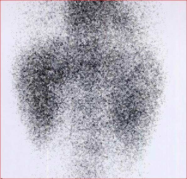
These pictures can help doctors assess how well the kidneys are working. DMSA travels through the body joined to a radioactive chemical. It builds up in the kidneys. Pictures of the kidneys are then taken using a special camera which can detect the radioactive chemical.
What is a DMSA scan used for?
This scan is used to check the structure of the kidneys, their size and shape. It is often used in children who have had unusual or repeated urinary tract infections.
It shows which areas of the kidney are working well and any areas of scarring. Scarring can be caused by a condition in which urine travels back from the bladder to the kidneys. This is called vesico-ureteric reflux. DMSA scans can also look for damage following an injury or reduced blood supply to the kidneys.
A DMSA scan enables doctors to see the functioning tissue of your kidneys. This is because DMSA does not attach itself to areas of the kidneys that are damaged. Doctors can compare the function of each kidney to see if one kidney functions differently to the other. By performing regular DMSA scans they can monitor any changes to inflammation of the kidneys.
Outras investigações such as an ultrasound scan can show the size and shape of the kidneys but not how they are working. This is why a DMSA scan may often be recommended in addition to other kidney tests or scans.
DMSA scans are usually requested by hospital specialists (usually paediatricians - doctors who specialise in children). The results should be given by the hospital specialist who requested the test, not by the GP.
How does a DMSA scan work?
A DMSA scan is a type of radionuclide scan. A radionuclide (sometimes called a radioisotope or isotope) is a chemical which emits a type of radioactivity called gamma rays.
A tiny amount of radionuclide is put into the body, usually by an injection into a vein. The radioactive chemical is often coupled to another substance. This substance takes the radioactive chemical to the part of the body doctors want to see.
It is possible to make many different types of radionuclides. Different ones tend to build up or concentrate in different organs or tissues. So, the radionuclide used depends on which part of the body is to be scanned.
In this case the substance DMSA is used because it builds up in the kidneys. Cells which are most 'active' in the kidney will take up more of the DMSA. So, active parts of the kidney tissue will emit more gamma rays than less active or inactive parts.
Os raios gama são semelhantes aos raios X e são detectados por um dispositivo chamado câmera de gama. Os raios gama emitidos de dentro do corpo são detectados pela câmera de gama, convertidos em um sinal elétrico e enviados a um computador.
The computer builds a picture by converting the differing intensities of radioactivity emitted into different colours or shades of grey. For example, areas of the target organ or tissue which emit lots of gamma rays may be shown as red spots ('hot spots') on the picture on the computer monitor.
Áreas que emitem baixos níveis de raios gama podem ser exibidas em azul ('pontos frios'). Diversas outras cores podem ser usadas para níveis intermediários de emissão de raios gama.
What happens during a DMSA scan?
A DMSA scan is usually done in the nuclear medicine department of a hospital. The first part of the scan involves a small injection. This is usually into a vein in the back of the hand. Children being scanned may be asked to come to the scanning unit an hour or so before the injection. This allows staff the time to put special Emla® cream on the back of a child's hand. Emla® cream is a local anaesthetic that numbs the area to reduce the discomfort of the injection.
You may also be asked to provide a urine sample (specimen) to make sure you do not have an active urine infection at the time of the test. If there is an infection, or if you have had a very recent infection, the scan may need to be postponed. This is because infection can alter the results of the scan and make it unreliable.
After the injection there will be a delay, usually around two to four hours, before the scan takes place. This allows enough time for the DMSA substance to travel around the body and reach the kidneys. Some hospitals will ask you to have some water or go to the toilet during this time, as this may make the pictures clearer.
After the wait the gamma camera is used to take pictures of the kidneys. It can take approximately 45 minutes to take all the pictures. During this time you need to remain as still as possible. If you are taking a child to be scanned it might be useful to bring your child's favourite book or toy to keep them occupied.
The camera can be quite big and for some pictures it may come quite close to your child's tummy (abdomen). It may be helpful to explain this before the test. Parents are usually allowed to stay with their children throughout the test.
How to prepare for a DMSA scan
Your local hospital should give you information on how to prepare for the scan. If you are pregnant, think you may be pregnant or are breastfeeding you must let the hospital know before the scan. You may be asked to drink lots of fluids before you attend.
Some hospitals recommend that you avoid medicines containing certain substances before the test. Your doctor should be able to advise you on this.
What to expect after a DMSA scan
The radioactive chemical you receive is eliminated from your body through the urine over the 24 hours following the scan. For that reason, you should drink plenty of fluids and urinate frequently following the injection. How much fluid will depend on each individual but you should be well hydrated and, for an adult, this could be 3-4 glasses of water.
The colour of your urine won't be affected by a DMSA scan. However, as it contains the radioactive tracer, it is recommended that you wash your hands well after going to the toilet.
In the case of babies and youngsters in nappies who are having a DMSA scan, there will be a small amount of radioactivity in the urine and therefore on the child's nappy. The radiotracer will not affect the baby's skin; however, carers should wash the baby's bottom as normal and wash their hands thoroughly.
Cloth nappies need to be washed thoroughly and disposable nappies put in a plastic bag and sealed before being disposed of.
If you have contact with children or pregnant women you should let your doctor know. Although the levels of radiation used in the scan are small the doctor may advise special precautions. Your hospital will be able to give you advice on this.
Possible side-effects or complications from a DMSA scan
The term 'radioactivity' may sound alarming. But, the radioactive chemicals used in radionuclide scans are considered to be safe and they leave the body quickly in the urine.
The dose of ionising radiation that your body receives is very small. In many cases, the level of radiation involved is not much different to a series of a few normal X-rays. However:
As with any other types of radiation (such as X-ray), there is a small risk that the gamma rays may affect an unborn baby. So, tell your doctor if you are pregnant or if you may be pregnant.
The scan usually isn't done if you are pregnant, unless it's absolutely necessary and can't be delayed until after the pregnancy.
If you're pregnant and your child is having a DMSA scan, it's usually recommended to get another adult to help look after them for the first 24 hours after the scan, as a precaution - and to avoid any contact with your child's bodily fluids (wee, poo, and vomit) for that time.
Raramente, algumas pessoas têm uma reação alérgica ao químico injetado.
Teoricamente, é possível sofrer uma overdose quando a substância química é injetada. Isso é muito raro.
Understanding the kidneys
Renal tract
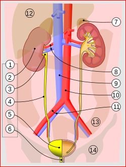
© Jordi March i Nogué [1], CC BY-SA 3.0, via Wikimedia Commons
There are two kidneys, one on each side of the tummy (abdomen). They make urine which drains down the ureters into the bladder. The ureters are tubes which go from each kidney to the bladder. Urine is stored in the bladder and, from time to time, is passed out through a tube called the urethra when we go to the toilet.
The kidneys, ureters, bladder and urethra are called the urinary tract.
Escolhas do paciente para Imagem

Exames e investigações
Exame DEXA
Exames de DEXA (também chamados de exames DXA ou densitometria óssea) são utilizados para verificar a densidade dos ossos. Este teste usa raios-X para mostrar quão fortes são os ossos. Um exame de DEXA é diferente de uma cintilografia óssea, que utiliza substâncias radioativas para criar uma imagem dos ossos.
por Dr. Colin Tidy, MRCGP
Exames e investigações
ressonância magnética
Uma ressonância magnética é um exame seguro e indolor que pode fornecer imagens detalhadas de órgãos e outras estruturas dentro do seu corpo. Nota: as informações abaixo são apenas um guia geral. Os procedimentos (e a forma como os exames são realizados) podem variar entre diferentes hospitais. Sempre siga as instruções dadas pelo seu médico ou hospital local. Estas geralmente estão incluídas na carta de agendamento.
por Dr. Toni Hazell, MRCGP
Perguntas frequentes
What is radionuclide imaging?
A DMSA scan is a type of radionuclide scan. A radionuclide is a chemical that emits gamma rays. A tiny amount of this chemical is injected into the body, usually into a vein, and is often linked to another substance that carries it to a specific organ or tissue. Different radionuclides can be made to concentrate in various organs, allowing doctors to target the part of the body they need to examine. In a DMSA scan, the DMSA substance carries the radioactive chemical to the kidneys.
How is the DMSA scan image produced?
The radioactive chemical taken up by the kidneys emits gamma rays. These gamma rays are detected by a special device called a gamma camera. The camera converts these rays into electrical signals, which a computer then uses to build a picture. The computer assigns different colours or shades of grey based on the intensity of the radioactivity; for instance, areas with high gamma ray emissions might appear as red 'hot spots', while areas with low emissions might be blue 'cold spots'.
Why is there a delay between the injection and the scan?
After the injection, there is typically a waiting period of about two to four hours before the actual scan begins. This delay is necessary to allow sufficient time for the DMSA substance, along with the radioactive chemical, to travel through the bloodstream and effectively build up within the kidney tissue. This ensures the kidneys are adequately 'lit up' for the gamma camera to capture clear and accurate images.
Will I feel unwell after a DMSA scan?
The radioactive chemicals used in DMSA scans are considered safe and are eliminated from your body relatively quickly through your urine, generally within 24 hours. The dose of radiation you receive is very small, often comparable to a few normal X-rays. While it's important to drink plenty of fluids and urinate frequently to help flush out the chemical, there are usually no significant side effects or complications like feeling unwell.
Are there any specific precautions for parents regarding contact with their child after a DMSA scan?
Yes, if you are pregnant and your child has a DMSA scan, it's recommended to have another adult help look after them for the first 24 hours as a precaution. During this time, you should also avoid contact with your child's bodily fluids (urine, faeces, and vomit). For babies and young children in nappies, there will be a small amount of radioactivity in the urine, so carers should wash the child's bottom as normal and wash their hands thoroughly afterwards. Disposable nappies should be sealed in a plastic bag before disposal, and cloth nappies should be washed thoroughly.
Why might a DMSA scan be postponed if I have a urine infection?
A DMSA scan might be postponed if you have an active urine infection, or if you've recently had one. This is because an infection can interfere with the results of the scan, making them unreliable. To ensure the accuracy of the test, and sometimes to avoid discomfort during the procedure, it's best to resolve any infection before proceeding with the scan.
Leitura adicional e referências
- Shaikh N, Spingarn RB, Hum SW; Dimercaptosuccinic acid scan or ultrasound in screening for vesicoureteral reflux among children with urinary tract infections. Cochrane Database Syst Rev. 2016 Jul 5;7:CD010657. doi: 10.1002/14651858.CD010657.pub2.
- Infecção do trato urinário - crianças; NICE CKS, abril de 2024 (acesso apenas no Reino Unido)
Sobre o autorVer biografia completa

Dr Doug McKechnie, MRCGP
Redator Médico
MA, MBBS, MSc, DRCOG, MRCP(UK), MRCGP(2021), FHEA
O Dr. Doug McKechnie é um médico do NHS que trabalha em Londres. Ele trabalha em tempo integral na prática clínica e também é o Vice-Líder do módulo de Prática Clínica e Profissional na Faculdade de Medicina da University College London.
Sobre o revisorVer biografia completa

Dra. Toni Hazell, MRCGP
MBBS, BSc, MRCGP, DFSRH, Dip GU med, DRCOG, DCH (London, UK, 2000)
A Dra. Toni Hazell se formou na Escola de Medicina do Hospital St. Mary e fez seu VTS no Hospital Northwick Park.
Histórico do artigo
As informações nesta página são escritas e revisadas por clínicos qualificados.
Artigo também disponível em Inglês, Alemão, Espanhol, Francês, Italiano, Português, Hindi, Hebraico, Árabe, e Sueco.
Próxima revisão prevista: 12 Nov 2028
14 Nov 2023 | Última versão

Pergunte, compartilhe, conecte-se.
Navegue por discussões, faça perguntas e compartilhe experiências em centenas de tópicos de saúde.

Sentindo-se mal?
Avalie seus sintomas online gratuitamente
Inscreva-se no boletim informativo do Patient
Sua dose semanal de conselhos de saúde claros e confiáveis - escritos para ajudá-lo a se sentir informado, confiante e no controle.
Ao se inscrever, você aceita nossos Política de Privacidade. Você pode cancelar a inscrição a qualquer momento. Nunca vendemos seus dados.
Mais em testes e investigações
- Audiologia
- Exames de sangue para detectar inflamação
- Bone scan
- Cateterismo cardíaco
- Enzimas cardíacas
- Teste de Coombs
- Eletrocardiograma
- Biópsia endometrial
- Teste imunológico fecal
- Hemograma completo e esfregaço de sangue
- Teste de tolerância à glicose
- Testes de audição
- Registro de pressão arterial domiciliar e ambulatorial
- Corpos cetônicos na urina
- Coleta de urina de meio de fluxo
- Teste de audição do recém-nascido
- Exames físicos para recém-nascidos
- Teste de contato para dermatite de contato
- Exame de sangue de rotina para função renal
- Verificador de sintomas