Pneumothorax
Trapped air in the chest
Peer reviewed by Dr Toni Hazell, FRCGPLast updated by Dr Philippa Vincent, MRCGPLast updated 13 Jan 2025
Meets Patient’s editorial guidelines
- DownloadDownload
- Share
- Language
- Discussion
- Audio Version
- Add to preferred sources on Google
In this series:Chest painCostochondritisBornholm diseasePleurisy
A pneumothorax is sometimes called a "collapsed lung" and it describes the condition in which air has become trapped next to a lung. Many cases occur without warning, particularly in healthy young men. Some develop as a complication of a chest injury or a lung disease.
The most common symptom is a sudden sharp chest pain followed by pains on breathing in. Some people become breathless. In most cases, the pneumothorax clears without needing treatment. The trapped air of a large pneumothorax may need to be removed if it causes breathing difficulty. An operation is needed in some cases.
At a glance
A pneumothorax occurs when air gets trapped between a lung and the chest wall.
This can cause a fully or partially collapsed lung.
Symptoms include sudden sharp chest pain and breathlessness.
A tension pneumothorax is a severe type needing urgent treatment.
A chest X-ray can confirm the diagnosis.
Small pneumothoraces may not need treatment, but larger ones require air removal.
Do not fly or scuba dive until a doctor confirms it is safe.
What is a pneumothorax?
A pneumothorax occurs when air becomes trapped between a lung and the chest wall.
Lungs are surrounded by a cavity called the pleural space. The pleural space is between the outside of the lungs and the inside of the chest wall. The pleural space normally contains a tiny amount of fluid (around 10mL) which helps the lung tissue to glide comfortably against the muscles as they inflate and deflate during breathing.
In a pneumothorax, air gets into this pleural space. Air in the pleural space can cause a fully or partially collapsed lung.
Lungs and airways - pneumothorax
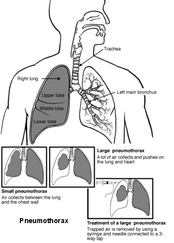
Types of pneumothorax
Spontaneous pneumothorax
A spontaneous pneumothorax happens suddenly without an obvious immediate cause. A spontaneous pneumothorax can be primary (occurring without any other lung condition) or secondary (occurring as a result of another lung condition).
Traumatic pneumothorax
A traumatic pneumothorax happens when the chest or lung is injured, allowing air to get into the pleural space. For example, a stab wound to the chest can cause a traumatic pneumothorax.
A traumatic pneumothorax can also happen as a complication of a medical procedure. For example, the procedure to insert a central venous catheter into the chest veins can sometimes accidentally cause a pneumothorax.
Tension pneumothorax
A tension pneumothorax is a severe and life-threatening type of pneumothorax. A tension pneumothorax can develop as a result of primary spontaneous, secondary spontaneous, or traumatic pneumothorax.
Tension pneumothorax causes shortness of breath that quickly becomes more and more severe. This occurs because the tear on the lung is acting like a one-way valve.
In effect, as each breath is taken in (inspiration), more air is pumped out of the lung and the valve action stops air coming back into the lung to equal the air pressure. The volume and pressure of the pneumothorax increases. This puts pressure on the lungs and heart. Emergency treatment is needed to release the trapped air; without urgent treatment, it can be fatal.
Pneumothorax symptoms
The typical symptom is a sharp sudden stabbing pain on one side of the chest.
The pain is usually made worse by breathing in (inspiration).
Breathlessness may occur, particularly in a large pneumothorax.
Other symptoms may exist if an injury or a lung disease is the cause - for example, cough or high temperature (fever).
A chest X-ray can confirm a pneumothorax. Other tests may be done if there is a suspicion of a lung disease being an underlying cause.
Pneumothorax causes
Primary spontaneous pneumothorax
This is where a pneumothorax develops for no apparent reason in an otherwise healthy person. This is a common type of pneumothorax. It is thought to be due to a tiny tear of an outer part of the lung - usually near the top of the lung.
It is not clear why this occurs but the tear often occurs at the site of a tiny bleb or bulla on the edge of a lung. A bleb or bulla is like a small balloon of tissue that may develop on the edge of a lung (a bulla is a larger version of a bleb). The wall of the bleb or bulla is not as strong as normal lung tissue and may tear. Air then escapes from the lung but gets trapped between the lung and chest wall.
Most primary spontaneous pneumothoraces occur in healthy young adults who do not have any lung disease. It is more common in tall thin people.
Secondary spontaneous pneumothorax
This means that a pneumothorax develops as a complication of an existing lung disease.
This is more likely to occur if the lung disease weakens the edge of the lung in some way as this may make the edge of the lung more liable to tear and allow air to escape from the lung.
For example, a pneumothorax may develop as a complication of chronic obstructive pulmonary disease (COPD) - especially where lung bullae have developed in this disease.
Other lung diseases that may be complicated by a pneumothorax include:
Other causes of pneumothorax
An injury to the chest - for example, a car crash or a stab wound.
Surgical operations.
Endometriosis - rarely, this can cause something called a catamenial pneumothorax.
How common is a pneumothorax?
About 2 in 10,000 young adults in the UK develop a spontaneous pneumothorax each year. Men are affected about three times more often than women and are affected at a younger age. Men are more likely to be affected around the age of 20 years and women in their early 30s.
It is more common in men who smoke than in men who don't smoke and nine times more common in women who smoke than in women who don't smoke. Cigarette smoke seems to make the wall of any bleb even weaker and more likely to tear.
Up to 5 in 10 people who have a primary spontaneous pneumothorax have another one or more at some time in the future. If it does occur again it is usually on the same side and it usually occurs within three years of the first one.
Complications of a pneumothorax
A pneumothorax can cause complications, such as:
Repeated episodes of pneumothorax in the future.
Infection in the pleural space (empyema, or pyopneumothorax).
Fluid inside the lung itself if it re-expands too quickly (re-expansion pulmonary oedema).
Bleeding into the pleural space (haemothorax, or haemopneumothorax).
In a tension pneumothorax, high-pressure air inside the pleural space can cause respiratory failure (inability to breathe) and ultimately a cardiac arrest (the heart stopping beating). A tension pneumothorax can be lethal if not treated urgently.
Diagnosing a pneumothorax
A pneumothorax can often be diagnosed by a doctor listening to the lungs. A chest x-ray will often confirm the diagnosis. Occasionally, other tests might be needed but this is unusual.
Pneumothorax treatment
No treatment
No treatment may be needed for a small pneumothorax. The small tear that caused the leak usually heals within a few days (sometimes as little as 1-3 days), especially in cases of primary spontaneous pneumothorax. Air then stops leaking in and out of the lung. The trapped air of the pneumothorax is gradually absorbed into the body.
A repeat chest x-ray may be needed in 7-10 days to check that it has gone. Simple painkillers might be needed for a few days if the pain is bad.
Removing the trapped air
This may be needed if there is a larger pneumothorax or if there are underlying lung or breathing problems. As a rule, a pneumothorax causing breathlessness is best removed.
The commonest method of removing the air is to insert a very thin tube through the chest wall with the aid of a needle. (Some local anaesthetic is injected into the skin first to make the procedure relatively painless.)
A large syringe with a three-way tap is attached to the thin tube that is inserted through the chest. The syringe sucks out some air and the three-way tap is turned. The air in the syringe is then expelled into the atmosphere. This is repeated until most of the air of the pneumothorax is removed.
Sometimes a larger chest tube is inserted to remove a large pneumothorax. This is more commonly needed for cases of secondary spontaneous pneumothorax when there is underlying lung disease. The tube is then left there for a few days to allow the lung tissue that has torn to heal.
Treating repeated episodes of pneumothorax
Some people have repeated episodes of spontaneous pneumothorax. If this occurs, a procedure may be advised with the aim of preventing the condition from coming back. For example, an operation may be an option to remove a bleb on the lung surface that repeatedly tears.
Another procedure that may be advised is for an irritant powder (usually a kind of talcum powder) to be put on the lung surface. This causes inflammation which then makes the lung surface stick to the inside of the chest wall.
A lung specialist will be able to give the pros and cons of the different procedures. The procedure advised may depend on the general health and on whether there is an underlying lung disease.
Preventing a pneumothorax
A smoker who has had a primary spontaneous pneumothorax can reduce the risk of its happening again by stopping smoking. Smoking tobacco, smoking cannabis, and vaping all increase the risk of a pneumothorax.
Flying, travel, and diving advice
It can be dangerous to fly with a pneumothorax. It is very important not to fly until a specialist has confirmed that it is safe to do so following a pneumothorax. This is usually around 7 days after a repeat X-ray has confirmed that the pneumothorax has disappeared completely.
Scuba diving increases the risk of developing a tension pneumothorax which can be fatal. People who have had a pneumothorax in the past are usually advised not to go scuba diving; they need a full evaluation by a private specialist in diving medicine if they still want to do so.
Patient picks for Chest pain

Chest and lungs
Precordial catch syndrome
Although precordial catch syndrome is a harmless cause of chest pain, there are many other causes of chest pain, some of which are serious and need urgent medical attention.
by Dr Colin Tidy, MRCGP

Chest and lungs
Chest pain
Chest pain refers to pain felt anywhere in the chest area from the level of your shoulders to the bottom of your ribs. It is a common symptom. There are many causes of chest pain. This leaflet only deals with the most common. It can often be difficult to diagnose the exact cause of chest pain without carrying out some tests and investigations.
by Dr Rosalyn Adleman, MRCGP
Frequently asked questions
What is the typical recovery time after a pneumothorax?
For a small pneumothorax, the tear that caused the air leak usually heals within a few days, sometimes as quickly as 1 to 3 days. The trapped air is then gradually reabsorbed by the body. A follow-up chest X-ray typically confirms its resolution within 7 to 10 days. If a larger chest tube was inserted, it might be left in place for a few days to allow the lung tissue to heal.
Can I prevent a pneumothorax from recurring if I've had one before?
If you are a smoker and have experienced a primary spontaneous pneumothorax, stopping smoking can significantly reduce the risk of it happening again. This includes avoiding tobacco, cannabis, and vaping, as they all increase the risk. Some people who have repeated episodes of spontaneous pneumothorax may be advised to undergo a procedure to prevent future occurrences, such as an operation to remove a weak spot on the lung surface or a procedure to make the lung surface stick to the chest wall.
Why are tall, thin people more prone to primary spontaneous pneumothorax?
Most primary spontaneous pneumothoraces occur in healthy young adults, and they are more common in tall, thin individuals. This type of pneumothorax is thought to be caused by a tiny tear in an outer part of the lung, often near the top, at the site of a small, balloon-like tissue called a bleb or bulla. The exact reason why tall, thin people are more susceptible isn't fully understood, but it's linked to these weaker areas on the lung surface.
What is a 'bleb' or 'bulla' on the lung?
A bleb or bulla is like a small balloon of tissue that can develop on the edge of a lung. A bulla is simply a larger version of a bleb. The wall of these blebs or bullae is not as strong as normal lung tissue, making them more likely to tear. When such a tear occurs, air can escape from the lung and become trapped between the lung and the chest wall, leading to a pneumothorax.
Are there any specific activities to avoid after a pneumothorax?
Yes, it is crucial to avoid flying until a specialist confirms it is safe, usually about 7 days after a repeat X-ray shows the pneumothorax has completely resolved. Scuba diving is also particularly risky because it increases the chance of developing a tension pneumothorax, which can be fatal. Individuals who have had a pneumothorax are generally advised not to scuba dive and should seek a full evaluation from a private specialist in diving medicine if they wish to pursue it.
Further reading and references
- British Thoracic Society Guideline for pleural disease; British Thoracic Society - BMJ (2023).
- Schnell J, Koryllos A, Lopez-Pastorini A, et al; Spontaneous Pneumothorax. Dtsch Arztebl Int. 2017 Nov 3;114(44):739-744. doi: 10.3238/arztebl.2017.0739.
- Goldman RD; Spontaneous pneumothorax in children. Can Fam Physician. 2020 Oct;66(10):737-738.
- Carson-Chahhoud KV, Wakai A, van Agteren JE, et al; Simple aspiration versus intercostal tube drainage for primary spontaneous pneumothorax in adults. Cochrane Database Syst Rev. 2017 Sep 7;9:CD004479. doi: 10.1002/14651858.CD004479.pub3.
About the authorView full bio

Dr Philippa Vincent, MRCGP
General Practitioner, Medical Author
MB BS, Bsc, MRCGP (2000), DCH, DFSRH, DRCOG
Dr Philippa Vincent is an NHS GP working in North London.
About the reviewerView full bio

Dr Toni Hazell, FRCGP
MBBS, BSc, FRCGP, DFSRH, Dip GU med, DRCOG, DCH (London, UK, 2000)
Dr. Toni Hazell qualified from St. Mary’s Hospital Medical School and did her VTS at Northwick Park Hospital.
Article history
Article also available in English, German, Spanish, French, Italian, Portuguese, Hindi, Hebrew, Arabic, and Swedish.
Next review due: 12 Jan 2028
13 Jan 2025 | Latest version
Join the conversation
Share your thoughts or experiences with others in our patient communities.

Ask, share, connect.
Browse discussions, ask questions, and share experiences across hundreds of health topics.

Feeling unwell?
Assess your symptoms online for free
Sign up to the Patient newsletter
Your weekly dose of clear, trustworthy health advice - written to help you feel informed, confident and in control.
By subscribing you accept our Privacy Policy. You can unsubscribe at any time. We never sell your data.