Eletroencefalograma
EEG
Revisado por Dr John Cox, MRCGPÚltima atualização por Dr Colin Tidy, MRCGPLast updated 24 May 2018
Atende aos diretrizes editoriais
- BaixarBaixar
- Compartilhar
- Language
- Discussão
- Versão em Áudio
- Add to preferred sources on Google
Nesta série:EpilepsiaTipos de convulsõesCrises tônico-clônicasCrises focaisCrises de ausênciaMedicação para epilepsia e efeitos colaterais
Esta página foi arquivada.
Não foi revisado recentemente e não está atualizado. Links externos e referências podem não funcionar mais.
An electroencephalograph is a useful test to help diagnose epilepsy. It records the electrical activity of the brain. However, a normal result does not rule out epilepsy.
Nota: as informações abaixo são apenas um guia geral. Os procedimentos e a forma como os testes são realizados podem variar entre diferentes hospitais. Sempre siga as instruções dadas pelo seu médico ou hospital local.
At a glance
An electroencephalograph (EEG) records electrical signals from your brain.
It is a painless and harmless test that usually takes 20-30 minutes.
Small patches (electrodes) are placed on your scalp and connected to the EEG machine.
An EEG can help diagnose conditions like epilepsy, especially if you have had seizure-like symptoms.
You may be asked to blink or breathe deeply during the test.
A normal EEG does not rule out epilepsy, and an abnormal result does not always mean you have epilepsy.
Neste artigo:
Video picks for Outros testes e investigações
Continue lendo abaixo
What is an electroencephalograph?
The brain normally produces tiny electrical signals that come from the brain cells and nerves which send messages to each other. These electrical signals can be detected and recorded by the electroencephalograph (EEG) machine. The EEG test is painless and harmless. (The EEG machine records electrical signals coming from your brain - it does not put any electricity into your brain or body.)
You may be advised to have an EEG if you have had symptoms which may be due to a seizure. (Old words for seizure are convulsion or 'fit'.) An EEG test is useful in diagnosing conditions such as epilepsy. See the separate leaflet called Types of Epilepsy and Seizures.
How is the test done?
Voltar ao conteúdoWhen you have the test, the operator will attach several small patches (electrodes) to your scalp. Wires from the electrodes are connected to the EEG machine. The machine detects and amplifies the electrical signals and records them on to a paper or computer. The test takes about 20-30 minutes. The electrodes are removed at the end of the test.
EEG machine
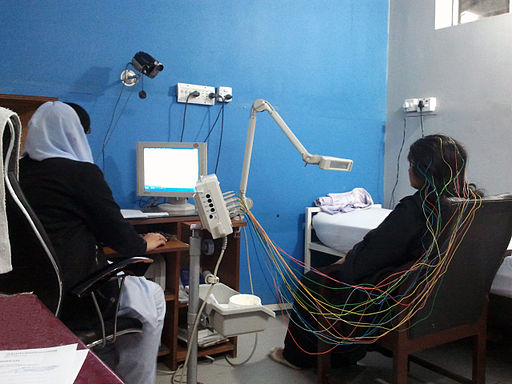
© By Baitaal (Own work), via Wikimedia Commons
For the duration of the test you may be asked to sit in a chair or lie on a couch. At some point you may be asked to blink lots of times, or to breathe deeply. These can sometimes trigger patterns of electrical activity in the brain which are associated with certain types of epilepsy.
Continue lendo abaixo
What can the test show?
Voltar ao conteúdoA normal ('negative') result
This shows a typical pattern of electrical activity from the brain. Most people without epilepsy, and many people with epilepsy, have a normal result. This is because an EEG only shows the electrical activity of the brain when the test is done. With many types of epilepsy, you only have abnormal electrical activity during a seizure. For the rest of the time, the pattern is normal.
An abnormal ('positive') result
This shows abnormal patterns of electrical activity. Some people with certain types of epilepsy have abnormal patterns all the time, not just when they have seizures. (Although, during a seizure the activity is even more abnormal.) For example, children with typical 'absence seizures' often have a characteristic EEG pattern which helps to confirm this type of epilepsy.
However, a small number of people who never have seizures and who don't have epilepsy, have some abnormal patterns of electrical activity in the brain.
Therefore, if you have symptoms which are thought to be seizures, an abnormal EEG means that the diagnosis is likely to be epilepsy. However, a normal result does not rule out epilepsy, and an abnormal result does not necessarily mean that you have epilepsy.
Children and electroencephalography
Voltar ao conteúdoThe interpretation of a child's EEG recording is more difficult. This is because the EEG changes during childhood. An adult pattern is usually developed by the age of 15 years. As the EEG pattern in infants and children can vary considerably, careful interpretation of the test is necessary.
Continue lendo abaixo
Some specialised types of electroencephalograph test
Voltar ao conteúdoStrobe lighting
In some cases, a strobe light may be used during an EEG test. This aims to detect if this alters the electrical pattern in the brain. (Usually it does not. However, a small number of people have seizures triggered by flickering or strobe lights and so this may help to identify these people.)
Sleep EEG
In this test, an EEG is performed while you are sleeping. This is usually carried out when you are in hospital. You may need to have one of these if your seizures happen when you are asleep or when you are tired.
Sleep-deprived EEG
There may be a better chance of detecting abnormal brain activity after a period of time when you are deprived of sleep. Therefore, sometimes the EEG test is done after you have stayed awake for all or most of the night. It is done in the same way as the normal test but with you asleep - after the period of 'sleep deprivation'.
Ambulatory EEG
This may be advised in cases where the diagnosis is not clear. It uses a portable EEG machine which records the brain's electrical activity when you are going about your normal activities. The electrodes can usually be hidden under your hair. The wires are connected to a small machine which you wear on a belt (a bit like wearing an mp3 player).
You may be asked to keep a diary when you have meals, go to sleep, and have any symptoms which may be a seizure. The patterns on the EEG can be analysed to see if they change when symptoms occur. It may help to confirm whether certain symptoms are due to seizures.
Video-telemetry
Where there is doubt about a diagnosis of epilepsy, or where the type of seizures someone experiences is unclear, video-telemetry can be helpful. This is a test that uses a video camera linked to an EEG machine.
The camera will visually record your movements and, at the same time, the EEG machine will record your brainwave pattern. Both the video and EEG are stored on to a computer so that they can be reviewed once the test is finished.
The doctor will be able to see any seizures that you may have had, as well as any changes in your EEG at that time. The test is often carried out over a number of days in order to increase the chances of recording one of your seizures.
What should I do to prepare for an electroencephalograph?
Voltar ao conteúdoYour doctor should give you instructions about what you need to do to prepare for an EEG. Commonly there is no preparation necessary. You should not stop any medication you take for seizures unless advised to by your doctor.
Patient picks for Outros testes e investigações

Exames e investigações
Aspiração por agulha fina
Na aspiração por agulha fina, uma agulha fina e oca é usada para coletar uma amostra de células de um órgão ou nódulo sob a pele. As células são então analisadas ao microscópio. Nota: as informações abaixo são apenas um guia geral. Os procedimentos e a forma como os testes são realizados podem variar entre diferentes hospitais. Sempre siga as instruções dadas pelo seu médico ou hospital local.
por Dr. Roger Henderson, MBBS

Exames e investigações
Teste do suor
Os testes de suor analisam a quantidade de sal deixada na pele quando uma pessoa transpira. Isso pode ajudar a diagnosticar fibrose cística. Nota: as informações abaixo são apenas um guia geral. Os procedimentos e a forma como os testes são realizados podem variar entre diferentes hospitais. Sempre siga as instruções dadas pelo seu médico ou hospital local.
por Dr. Colin Tidy, MRCGP
Perguntas frequentes
Will I feel anything when the electrical signals are detected by the EEG machine?
No, the EEG test is painless and harmless. The machine records the electrical signals naturally produced by your brain; it does not send any electricity into your brain or body.
What specifically happens during the test to try and trigger brain activity?
During the test, you might be asked to blink many times or to breathe deeply. These actions can sometimes help to trigger specific patterns of electrical activity in your brain that are linked to certain types of epilepsy.
If my initial EEG is normal, does that mean I definitely don't have epilepsy?
Not necessarily. A normal (negative) result indicates a typical pattern of electrical activity at the time of the test. However, with many types of epilepsy, abnormal electrical activity only occurs during a seizure. For the rest of the time, the brain's electrical pattern can appear normal. Therefore, a normal result doesn't rule out epilepsy.
What if my EEG shows abnormal patterns, but I don't have seizures?
It is possible for a small number of people who never have seizures and do not have epilepsy to show some abnormal electrical patterns in their brain during an EEG. Therefore, an abnormal result doesn't automatically mean you have epilepsy.
How does an ambulatory EEG work if I'm moving around?
An ambulatory EEG involves a portable machine that records your brain's electrical activity as you go about your daily activities. The electrodes are usually hidden under your hair, and they are connected to a small machine worn on a belt. You might be asked to keep a diary of your meals, sleep, and any potential seizure symptoms to help analyse the patterns recorded.
Why would a video-telemetry test be recommended?
Video-telemetry is useful when there is uncertainty about an epilepsy diagnosis or if the specific type of seizures someone experiences is unclear. This test combines a video camera, which records your movements, with an EEG machine, which records your brainwave pattern simultaneously. This allows doctors to match visual seizure events with changes in your brain's electrical activity. It's often conducted over several days to increase the chance of recording a seizure.
Leitura adicional e referências
- Epilepsias: diagnóstico e manejo; Diretriz Clínica do NICE (Janeiro de 2012)
- Diagnóstico e manejo da epilepsia em adultos; Rede Escocesa de Diretrizes Intercolegiais - SIGN (2015 - atualizado 2018)
- Epilepsia; NICE CKS, março de 2018 (acesso apenas no Reino Unido)
Continue lendo abaixo
About the authorView full bio

Dr Colin Tidy, MRCGP
Médico Generalista, Autor Médico
MBBS, MRCGP, MRCP (Paediatrics), DCH
Dr Colin Tidy é um médico do NHS, baseado em Oxfordshire.
About the reviewerView full bio

Dr John Cox, MRCGP
MA, MB, B Chir, DCH, DRCOG, MRCP (UK), MRCGP
Dr John Cox worked as a Medical Registrar in the UK and New Zealand and as a locum Physician in New Zealand.
Histórico do artigo
As informações nesta página são escritas e revisadas por clínicos qualificados.
24 May 2018 | Última versão

Pergunte, compartilhe, conecte-se.
Navegue por discussões, faça perguntas e compartilhe experiências em centenas de tópicos de saúde.

Sentindo-se mal?
Avalie seus sintomas online gratuitamente
Inscreva-se no boletim informativo do Patient
Sua dose semanal de conselhos de saúde claros e confiáveis - escritos para ajudá-lo a se sentir informado, confiante e no controle.
By subscribing you accept our Política de Privacidade. Você pode cancelar a inscrição a qualquer momento. Nunca vendemos seus dados.